Introduction:
While there are myriad exotic life forms that can be encountered in a single drop of pond water, actually finding and seeing them can be a challenge. This page provides hints and techniques for capturing the 17 most common freshwater pond creatures. The following images and videos are what you can expect to see and photograph using the pedestrian microscopes commonly available to the public for $200 - $1000. While these can provide hours of fun at low power, they simply don't have the optical quality to provide sharp images much above 200X. The extraordinary videos available on-line from professional microphotographers like Mr. Craig Smith (search youtube for "craig smith protists" for stunning examples) are achieved using the world's best optical microscopes from Nikon, Olympus, Leica or Zeiss that can crowd $20,000 with all the bells and whistles.
The secret to collecting as many different types of microscopic organisms as possible is to avoid crystal clear flowing water and instead let your nose lead you an area of very still water heavily overgrown with string or hair algae.
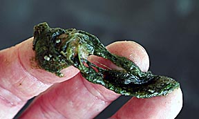
Just as ocean kelp fosters a wide diversity in fish life, so string algae promotes microscopic life.
Avoid loose, clumping algae that floats in drifts on the surface of ponds. This does little more that fill the microscope's field of view with dark blotches and doesn't harbor the amount and diversity of life that string algae offers.
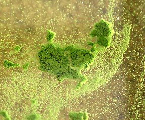
Clump algae
Take samples from as many different locations as possible: as far under the surface as you can reach, from the surface itself away from shore, in brightly sunlit zones as well as those in shade, scoop up sand from the bottom and collect water from the the shallowest spots near the shore. Also, visit as many different lakes, ponds and even drainage ditches as possible. Each has it's own unique biosphere.
Mix the samples together in a holding tank. I prefer a ten gallon aquarium equipped with a bubbler and a light at one side to attract microorganisms.
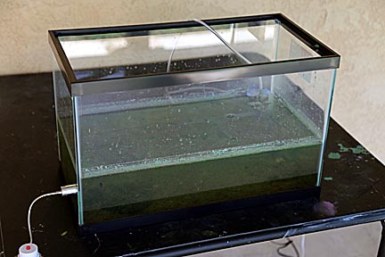
Most importantly: Keep this tank outside and away from windows! It's odor can be strong enough to take away your breath. The bubbler will help maintain oxygen levels in the water, which will prevent some of the purtification that always accompanies green tanks like these, but won't stop all of it.
When preparing samples, use an eyedropper and bring only the smallest quantities into the house.
It's a good idea to wear plastic gloves to protect your hands when collecting samples. It's possible to come in contact with some life forms that can be dangerous.
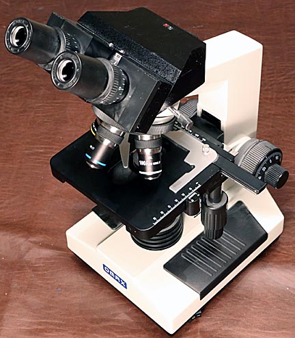
My inexpensive $400 OMAX Binocular Microscope with built in video camera.
If this is going to be your first time using a microscope be prepared for an exercise in frustration. Even the best models have optical limitations that make viewing the smallest object all but impossible.
The problem is that microscopes aren't designed to view three-dimensional objects like the living microbes we'll be hunting. Rather, they work best on very thin, flat slices of objects. The problem with 3-D objects is that microscopes have incredibly short depths of field.
Depth of field is the distance over which an object appears to be in sharp focus. Let's say you're looking at a water flea that's 2 millimeters thick. You focus on it's eye, which is halfway between the flea's far side and close side - the side facing you. Even at the microscope's lowest power, typically 40X, the depth of field will only be 0.5 mm. That means anything closer or further away than 0.25 mm from the eye will be out of focus. Worst still, these out-of-focus zones will overlay the in-focus areas and make them harder to see. This problem increases as magnification increases. It's a property of optical physics and has nothing to do with the quality of the microscope.
Compounding this problem is the quality of the microscope. Cheap scopes have poorly corrected optics that may exhibit chromatic aberrations. Just like white light passing through a prism splits into a rainbow, so light passing through a lens separates into different colors. These present themselves as colored bands surrounding the object. Because most microbes are transparent to some degree, these colored rings surround all internal and external structures in overlapping rainbows that blur the image.
Finally, the microscope has to deal with diffraction. Because light acts like a wave, as it passes through a small aperture, like a microscope's lens or the light source's adjustable aperture, bright and dark rings are created. Just as with chromatic aberrations, these further weaken the image.
Having read the above you might be thinking it's impossible to get any sort of viewable image out of a microscope. Yet with a few simple techniques you can. First, even if the microscope is capable of reaching 2000X, assume the maximum useful magnification is 100X. Second, after placing a drop of sample water on a slide, always cover it with a thin cover slide. This will confine microbes to a thin enough plain to be mostly in focus. Third, forget about going after the smallest microbes. You'll often see tiny specks swimming around at low power. To magnify them sufficiently to see any detail you'd have to use such high magnifications that optical distortions will blur them to unrecognizability. Finally, stop the light source's aperture down as small as possible to maximize the depth of field.
One other issue is the fact that most of the living organisms you'll be observing move so fast that they are all but impossible to keep in view. There are chemicals sold by microscope supplies that can be added to the water to slow them down.
If you haven't purchased a microscope yet, I highly recommend you do so only after finding a retailer who'll let you look through it. My OMAX came from Amazon.com and while the reviews provided some indication it was a good instrument, I soon discovered several problems. The video camera has an extra magnification factor so that the view through it is not the same as through the eyepiece. At 40X the eyepiece provides a field of view that's 4.5 mm across. At the same setting the view through the camera is only 2.5 mm, suggesting it's operating at 75X. The focus is not the same for the eyepiece and camera. The camera's frame rate is only four per second, which results in jerky movement. These are things that would be extremely difficult to discover without being able to take it for a test drive.
Something that can't be emphasized enough is that all the images and videos on this page were taken through the camera, which greatly reduces the sharpness. Through the eyepiece, these microbes show much more detail and color.
Once you have gotten pond water and a microscope, it's time to have some fun. Taking samples samples from deep within a lump of string algae, the clear water in the middle of the green tank or from the sand laying on the bottom of the tank will provide different types of microorganisms. The following seventeen are the most common:

Daphnia, or
water fleas, range from 1 to 4 mm across and can be seen with the
naked eye.
They are
crustaceans related to shrimp and eat anything that sweeps into their mouths.
Often they
will have a bright green ribbon running through them from the algae
they've consumed.
Viewed best at
the lowest power.
Amphileptus is
a tiny predatory worm that's barely 0.05 mm long.
Viewed best at 100X.
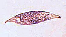
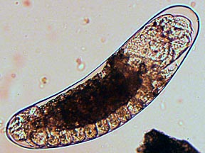
This flatworm,
possibly rhabdoceola, is the same size as amphileptus, but viewed
here at 400X.
Note the two
eye spots.
See the
diffraction shadow around it?
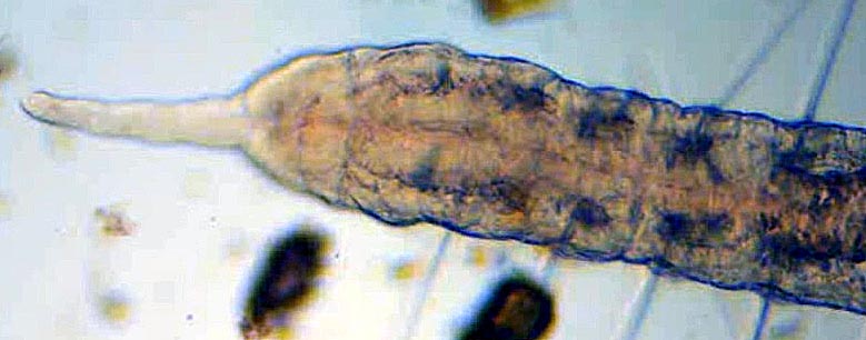
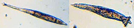
Flatworms vary
greatly in size. Compare the one about at high power to the following
at only 40X... and this is just it's head:
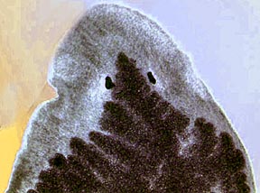
This guy measured 6 mm from nose to tail.
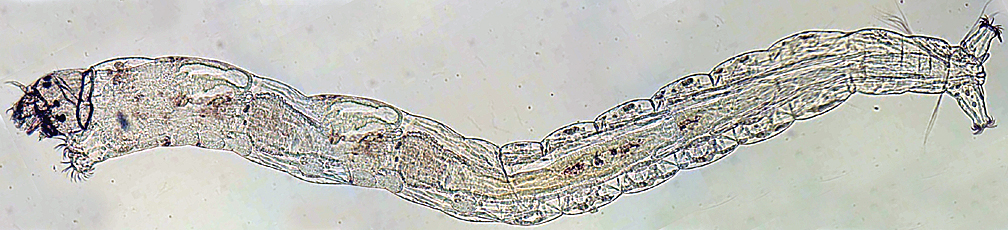
Wandering
around a drop of water with a microscope while looking for tiny
microbes can result in surprises, such as when you come across a
large worm or larva.
This is just the head of an Oligochaster Diastrophus at 40X. Altogether he stretched 8 mm.
A 7 mm long midge fly larva. This image was assembled by combining five exposures.
Other surprise discoveries pop up when you least expect them, like the following example of what I at first thought was a new type of blue ribbon algae:
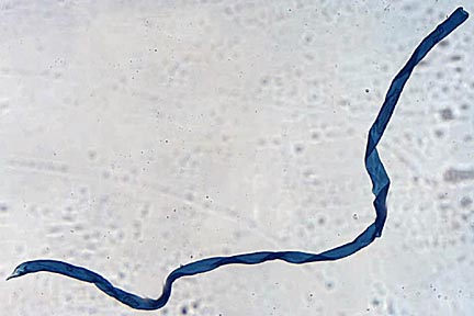
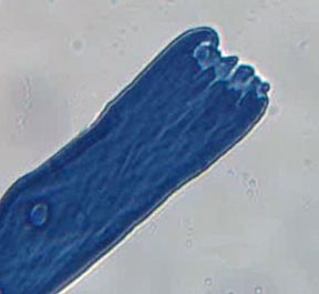
Then I sneezed and it disappeared. My great discovery turned out to be a bit of lint that had landed on top of the slide.
Let's get back to the small stuff!
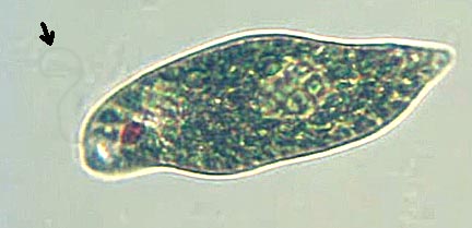
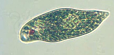
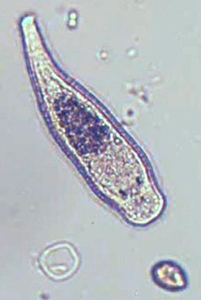
This is
Euglena, a single celled microbe that can both eat and has
chlorophyll like a plant. Note the red eye spot.
The arrow
points toward an almost invisible hair it whips around to propel itself.
Viewed best at 100X.
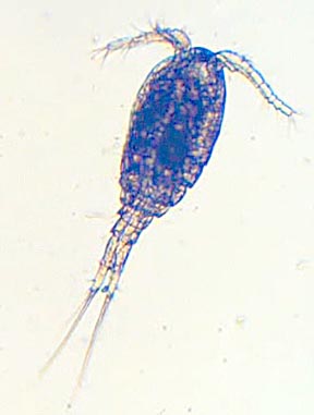
Cyclops, so
named because it has only one eye.
Viewed best at
100X but easy to spot at lower powers.
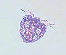
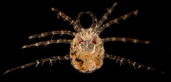
This little
guy is a nauplius, the larva of a microscopic crustacean.
I found I
could see the most detail at 100X.
From the sand,
this pupa could hatch into almost anything.
40X.

I believe this might be a small paramecium, but at 1000X the details are so distorted it's impossible to say.
Rotifers are
multicell predators that sweep food into their mouths by whirling
tiny hairs called cilia on their heads.
They have two
fingers on their tails, used to hold onto a base. They move either by
inching
forward like an inch worm or using their cilia to pull them rapidely through
the water.
Viewed best at 100X but pushing the magnification to 400X
provides a
hint of the moving cilia.
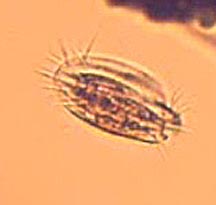
Tiny Hypotrich is needs 400X to bring out any detail. At 40X they are just little dots zomming around.
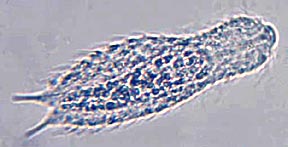
A Gastrotrich
Chaetonutus, a multicelled predator, looks like a termite wandering
around the slide.
He looks best
at 100X. At 400X, like this image, optical problems hide as much
detail as the extra power reveals.
The following images show a few of the many forms algae can take:
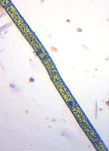
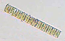
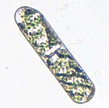
The next two objects are diatoms, algaes with hard transparent shells. They slowly push themselves around:
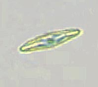
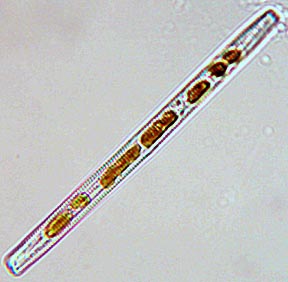
The shells of dead diatoms are what make up the diatomaceous earth used in swimming pool filters.
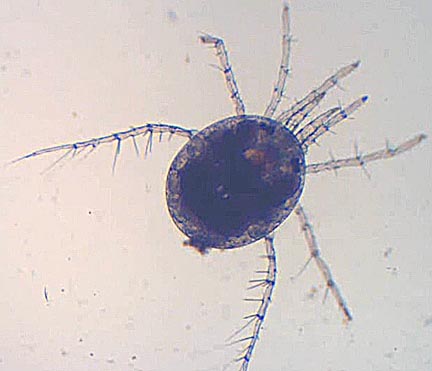
This 1 mm diameter spider is actually a water mite. Best viewed at the lowest possible magnification.
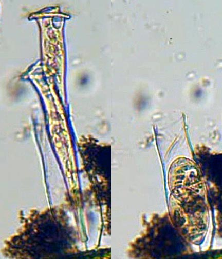
This is
vaginicola, a trumpet shaped ciliate which, like rotifers, use cilia
to pull food into their mouths.
Unlike
rotifers, these peritrich ciliates are single celled microbes. The
left image shows them feeding.
When disturbed
they retract into a transparent protective shell called a lorica, as
seen on the right.
Vaginicola is
distinguished from other loricated ciliates by the symbiotic green
algae in its body.
The smaller
ones can most easily be spotted by looking for small pieces of debris
whirling in
circles. Average sized specimens are viewed best at 100X.
Stentors are larger versions of paritrich ciliates like the above, except that they have not loricas and can grow to be 2mm long.
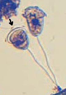
Vorticella are
bell shaped cousins of Stentors. Tethered to a base by a long thread,
they jerk back when disturbed.
The arrow
points to one of the cilia waves moving around its mouth. Viewed best
at 100X.
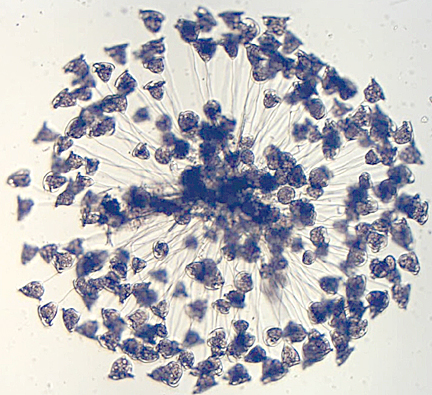
Undoubtedly the most magnificent object to be seen is a spherical colony of vorticella, which can measure over 1 mm in diameter. Their constant retractions make them look like a glorious sea urchin.
This is one
object you don't want to view with a cover slide because it'll crush
the sphere.
Instead, set
your microscope's magnification to minimum and leave the drop mounded
on the slide.
The following video shows what these microbes look like in live action:
How to Convert a Brightfield Microscope to a Darkfield Microscope:
The easiest and cheapest microscope modification that yields the greatest rewards is converting it to a darkfield instrument. To do so, cut a clear plastic disc the same size as your filter holder. Cut a circle out of black vinyl tape and mount it in the center of the disc. The size will vary depending on the microscope. For my OMAX, a black disk 17 mm in diameter worked well for 40X and 100X. For higher powers, either use a higher power eyepiece or purchase a special, and unfortunately expensive, high power darkfield condenser.
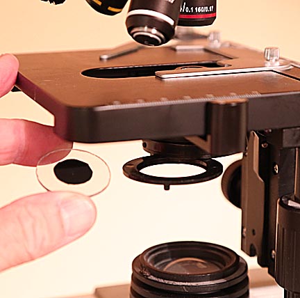
Place the darkfield adapter in the filter holder, rotate it into the light path and instead of images like this:
You'll see something like this:
What's happening is that the black circle blocks the light going straight into the objective, which is what creates the bright background you usually see. The object under observation is illuminated by peripheral light passing through the clear outer area of the disk. Cast against a dark background, the object has much greater contrast making it possible to see more detail.
The one drawback is that dust on lenses and slides are also more easily seen, which sometimes makes identifying objects more difficult.

And speaking of identification, with over known 50,000 microbes it can be very difficult to find out what you are looking at. The protist above, which I named "the corkscrew" is such an example. An hour's research on the internet failed to provide information on what it was. The reason is that because a microbe's appearance varies greatly with viewing angle and technique and the images in many references are so poor, figuring out what you're looking at can be extremely problematic. I found two of these and neither moved. However, for all I could find out they could be dead vorticella with their anchor threads curled up into a spring shape.
Still, darkfield microscopy is well worth the small investment in time a resources. The following video shows a few of the microbes I recorded with my system:
For someone with an average hobby microscope, typically costing a few hundred dollars, identifying microbes with certainty is a frustrating challenge, as the following video shows:
Conclusions:
The microscopic world offers some of the most hideous and most beautiful life forms to be seen on Earth. With a microscope and a little patience even a casual observer will be able to discover unimaginable wonders. I sincerely hope this page has whetted your interest in this fascinating hobby.

Return to my main page to browse 60 other subjects